

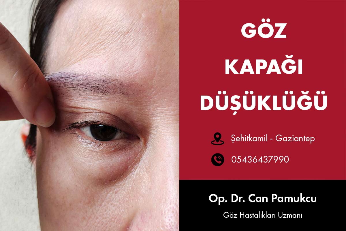
Impairment Of The Eyelid
Contents
- Impairment Of The Eyelid
- Causes Of Low Levels Of The Eyelid
- Clinical Appearance Pitozis
- Ptosis Examination
- Eye Examination
- Ptosis Private Examination
- What Are The Types Of Ptosis?
- Nov Connected To Decomposition Ptosis
- Neural Ptosis
- Third Cranial Nerve Palsy
- Horner's Syndrome
- Nov Ptosis Disease
- Myasthenia Gravis
- Myotonic Dystrophy
- Chronic progressive External Ophthalmoplegia (CPEO)
- Nov Okulofaringeal Dystrophy
- Mechanical Ptosis
- Traumatic Ptosis
- Psodoptozis
- Treatment Of Eyelid Impairment
- Results
Causes Of Low Levels Of The Eyelid
Impairment of the eyelid as a result of several factors may be innate in each age group, as can occur and may not occur. Patient ptosis if they are applied with a complaint, this is not a diagnosis, a condition that may occur as a result of another disease, it should be noted that. A comprehensive assessment and examination to determine the cause is extremely important.
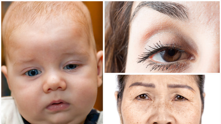
Innate reasons: In some babies, the muscle that allows the eye lid to be removed due to a lack of congenital ptosis can be seen.
Age-related changes: As you get older, as a result of the weakening of the muscle that lifts the eyelid ptosis may occur.
Trauma: Received from the impacts of eyelid ptosis can occur as a result of.
Neurological causes: Some nervous disorders can lead to the degradation of the eyelid.
Clinical Appearance Pitozis
Patients with pitozi usually suffer from the following problems.
- Sagging of the eyelids
- Feeling of heaviness in the eyes
- Visual blurring due to sagging
- Cosmetic complaints
A thorough history taking and clinical examination, the identification of the etiology of pitoz and helps you to plan the appropriate treatment.
Getting the story, pitoz of starting age, progression, duration, and aggravating or mitigating factors should include. Double vision, Daily variability, pain, eyelid swelling, or difficulty swallowing, such as temporary weakness of the symptoms associated with the diagnosis helps make Nov. Trauma, ocular or eyelid surgery, contact lenses, and predisposing factors such as botulinum toxin injections should be carefully excluded.
To exclude inherited disorders in the family history of ptosis should be sought. The story of old photographs, some patients the disease gives an idea about the starting time of the evaluation.
Any systemic illness, mental health problems and drug requires documentation of history.
Medication to patients who are using blood thinners such as Aspirin, before the drugs are recommended to leave 1 week after surgery.
Ptosis Examination
Clinical examination, the patient begins from the moment you entered the doctor's clinic. Any facial asymmetry, excessive movement that connects, chin tilted up to look at whether or upside posture is important.
Eye Examination
- Visual acuity and refraction time
- Cover and test component to exclude any call you any hipotropi psodoptozi
- Any abnormal extraocular motility disorder and eyelid movement
- Horner's syndrome or 3. a pupil for examination cranial nerve paralysis
- Giant papillary conjunctivitis or examine to look for any semblefaron
- Corneal sensation and dry eye assessment may increase susceptibility to postoperative keratopati.
- Fundus examination, Retinal pigmentary degeneration of the properties for
Ptosis Private Examination
Measurements of the eyelid muscle that connects the position of the eyes with the thumb straight and prevent movement should be made in view. The person who examined the patient at eye level to prevent the error should be seated.
1) Palpebral fissure height (PFH): Overview and position in the pupil plane vertical palpebral aperture height between the edge of the upper and lower eye lid. The average distance is close to 10 mm.
2) Marginal reflex Distance 1 (MRD 1): MRD 1, the corneal light reflex is the distance between the top cover edge. Normal MRD 1, 4-5 mm. 1 MRD difference between two eyes, one-sided pitoz pitoz of patients with mild, moderate or severe to be classified as helps.
The difference between the two eyes in the MRD 1:
- 2mm – mild sagging
- 3mm – moderate sagging
- 4mm – Severe ptosis
3) Marginal reflex Distance 2 (2 MRD): 2 MRD, with the corneal light reflex is the distance between the edge of the lower eyelid. Normally MRD 1 + 2 MRD = eye cover range
4) Levator movement: Excessive excessive muscle movement by blocking the lifting of the eyelid excursion measured with millimeter scale down to look up from the look during the moving process. The lifting movement is 15 mm larger than normal. Pitozisli the selection of the surgical procedure in a patient is the most important as it determines the value of the measurement.
Leverage the grading of movement
- Less than 4 mm – Bad
- 5 to 9 mm – Medium
- 9 to 11 mm – good
- Larger than 12 mm – Excellent
The lifting movement is weak (less than 4 mm) in patients the love that connects surgery the preferred process.
5) The curvature of the edge distance (MCD)Glance down at the edge of the cover is the distance between the folds of the skin. MCD Normal 7 to 8 mm in men and 8 to 10 mm in females. Whereas in MCD MCD is usually congenital or dimmed in pitoz aponorotik pitoz is no higher than usual.
To provide and maintain a good aesthetic eye during surgery with the same symmetry against the realignment of curves is very important.
6) Bell's phenomenonThis is a very important factor that needs to be considered before the correction of pitoz. The patient's eyes slowly prompted to shut down, and tries to open it. This reflex in patients who are weak after surgery to avoid the risk of exposure to keratopati should be avoided or corrected is less than required for the maximum amount of pitoz should be corrected.
7) if you have that is going to make things worse after the surgery, and eyelid lagoftalmi glance down at the presence of the delay should be evaluated.
8) if you have Nov, low or sagging in the skin of the upper lid must be recorded. Also involusyonel ptosis upper lid surgery process often it is combined with ptosis repair.
9) Test Hering: Single-sided pitozlu in patients pitotik cover gently removed manually, and the contralateral eyelid is observed. Due to the law of equal innervation Hering, may be reduced to cover the other eye (Seesaw effect). Show ptosis requiring surgery in this patient prior to surgery and warn them about the possibility of the opposite eye is important. In such cases, treatment may be missing a planned fix.
10) Oxycodone testMild ptosis, is a useful test in patients with Horner's syndrome or linked to pitozisli; fornik if the top 2.5% phenylephrine drops is dripping. Ptosis measurements are repeated after 10 minutes. The higher the number of patients due to the stimulation of Müller muscle posterior approach with correction of eyelid pitotik pitoz (konjonktival – mullerektomi are ideal candidates for surgery.
11) Tests to rule out myasthenia gravis:
Fatigue test: The patient maintains fixation for 30 seconds at a glance. Nov Miyastenili patients due to fatigue eyelids slowly falls down.
Ice test: An ice pack placed over the closed eyelid for 2 minutes. Ptotik measurements are repeated after 2 minutes. Pfh 2 mm or more is considered positive for myasthenia gravis in healing. The reason for this is to improve neuromuscular transmission cooling.
12) Ekzoftalmometri HertelA Hertel read, or you exclude to the exclusion of any proptoz enoftalm psodoptozi and hence helps helps.
What Are The Types Of Ptosis?
Impairment of the eyelid, ptosis is classified as true, false and fair. True ptosis, ptosis ptosis is classified as congenital and acquired by the age of occurrence.
In addition to the etiologic factors are classified pitoz adult acquired:
- Nov connected to decomposition ptosis
- Neurogenic ptosis so nervous
- Nov myogenic ptosis diseases due to so
- Namely mechanical, scratching, picking, ptosis due to wrong makeup techniques
- Traumatic ptosis due to an accident
Nov Connected To Decomposition Ptosis
Ptosis Aponevrotik ptosis is the most common form of adult and usually 50 – 60 years old occurs. However, trauma in young individuals, recent eyelid swelling, eye surgery or long-term contact lens use after may occur.
The pathogenesis of the muscle that lifts the eyelid aponevrotik carrying mostly stems from the separation of the upper lid. Age-related cases, there is separation real, and sometimes due to stress or the thinning of aponevroz ptosis occur. The concentration of fat in the muscle that lifts the eyelid sometimes seen.This type pitoz the characteristics of patients with good levator function to be equipped with high cover of the folds, the affected eyelid upper eyelid excess skin is thin with a glance down and look below.
Neural Ptosis
Neurogenic ptosis the muscle that lifts the eyelid is caused by any situation that disrupts or below the muscle of Müller. 3 the most common varieties of an eye doctor. is Horner's syndrome and cranial nerve palsy.
Third Cranial Nerve Palsy
Lesions that occur along the oculomotor nerve, and adduction of the eyeball pitozi, elevation, and by restricting the movements of depression manifests itself. Pupil involvement may or may not be present. Bell's phenomenon is usually weak.
Third nerve palsy was caused by artery aneurysm is considered to be mostly for neurological compress the nerve. Third nerve palsy is mostly caused ischemic vascular protective Pupil and usually will clear up within 3 months. Among other reasons along the border inflammation, trauma, or tumors in the lobby. Upper orbital fissure cranial nerve palsy seen with the other branch originates the orbit or cavernous sinus lesions.
Due to the presence or absence of Bell's phenomenon in patients with weak after surgery because of the risk of exposure to high keratopati difficult to treat. Ideally, before strabismus surgery is performed to correct the deviation, then fix with planned missing suspension technique that connects with ptosis correction.
Horner's Syndrome
Horner's syndrome mild ptosis, pupil and myosin consists of anhidroz enoftalmi significant. Müller muscle and pupillary dilator muscle sympathetic nerve occurs due to the discontinuation of supply.
In dim light the pupil anisocoria can be shown in a good way. Emerging in childhood in patients with Horner's syndrome, sympathetic eye darkening melanin production in melanocytes which are controlled by the wanes as the way there.
The diagnosis of Horner's syndrome is often made clinically. 4 %cocaine %1 hidroksiamfetamin or 2.5% phenylephrine made using pharmacological tests help confirm the diagnosis.
Nov Ptosis Disease
Myogenic ptosis, the levator muscle due to an abnormality in itself will occur. These patients are usually limited mobility and decreased extraocular movement, facial expressions, they levator in conjunction with the resort.
Myasthenia Gravis
Myasthenia gravis, antibodies against acetylcholine receptors in the neuromuscular endplate of the voluntary muscles is an autoimmune disease characterized by the presence of. This causes weakness and fatigue leading to a decrease of the effect of acetylcholine by opening Nov. Myasthenia gravis may be generalized or localized to the Eye (Ocular myasthenia).
Double vision is the most common pitoz variable associated with the property. Symptoms may be unilateral or bilateral. For usage in myasthenia, most patients initially has the function of a good cover Nov. Long-term-up overview, due to fatigue in these patients will lead to a deterioration of pitoz Nov.
Other tests that help confirm the diagnosis between the ice test, serum acetylcholine receptor antibody test, repetitive nerve stimulation and single fiber test elektromiyografi facility.
The treatment of such patients acetylcholinesterase drugs, or immunosupresan includes the implementation of oral steroids. In patients with severe pitozlu Fix missing pitoz planned correction may be an option.
Myotonic Dystrophy
Myotonic dystrophyslowly progressive ptosis and external ophthalmoplegia that occur with an autosomal dominant disease. Pathological processes, muscle contraction after is to relax. At the same time on the face, neck and limb muscles included.
These patients also disperse light close to the pupil eye examination, chromatic cataracts (cataract and retinal pigmentary degeneration Christmas tree are also seen. Develops a model of testicular atrophy in men androgen and a frontal.
Chronic progressive External Ophthalmoplegia (CPEO)
CPEO, bilateral symmetrical involvement of the extraocular muscles is a disease that causes a mitochondrial Nov. Symptoms in adulthood begins in childhood or adolescence and progresses slowly. The initial symptom is bilateral symmetrical involvement, and this is followed by a two-sided mutation.
Due to the symmetrical involvement of the muscles of the iris, patients often complain without seeing double. In addition to including the muscles of facial expression, expressionless face develops. (Hutchinson side)
Diagnosis is confirmed by biopsy due to enlarged mitochondria with irregular red fibers seen Nov.
Kearns-Sayre syndrome: Retinal pigment degeneration, cardiac conduction defects, complete heart block, ataxia, neuropathy, endocrine dysfunction that occurs in young adults showing a CPEO and varieties.
CPEO is not available for any treatment until today. In severe cases of exposure to high risk by keeping in mind that keratopati to clear the visual axis impairment eyelid surgery can be done.
Nov Okulofaringeal Dystrophy
This autosomal dominant disorder, life of between 4 and 5. within a decade, bilateral ptosis, progressive external ophthalmoplegia, dysphagia, dysarthria, facial weakness in the muscles and proximal limb weakness manifests itself.
Mechanical Ptosis
Of the valve to increase the weight, or any tumor that causes ptosis secondary to scar formation and wound in the conjunctiva blefarosalaz.
Traumatic Ptosis
Ptosis occurs due to direct or indirect trauma to the levator muscle. The muscle that lifts the lid by blunt force trauma and penetrating injuries after including ptosis may develop. Wait 6 months and then get better after trauma fair that can be surgically repaired.
Psodoptozis
This is not a real ptosis, the levator muscle is caused by abnormalities in a significant pitoz outside of the structure.
Among the reasons dermatosalazis, Nov impairment, hipotropi, revealed anophthalmia, and contralateral eyelid retraction fitizis ampuli takes place.
Before the surgical correction of excess skin that distinguish it from the real pitoz psodoptozis is very important.
Treatment Of Eyelid Impairment
Treatment of eyelid impairment, may vary depending on the cause and severity of ptosis. Surgery, physical therapy or drug therapy, such as different approaches can be used.
Results
Ptosisaesthetic and functional can lead to problems. Early diagnosis, it is important to avoid potential complications and determine the most effective treatment method. If you're suffering from this issue you also, you can find out the most appropriate treatment method by referring to an eye specialist.